
The Case for Multidisciplinary, Individualized Care in AVM Treatment

Even as a healthy 49-year-old nurse, Sharron Fowler found herself trapped in her own medical mystery. Experiencing pain, balance issues, and difficulty thinking, she knew something was off. She was admitted to the hospital where she worked, but after a few days and no clear diagnosis, she was discharged.
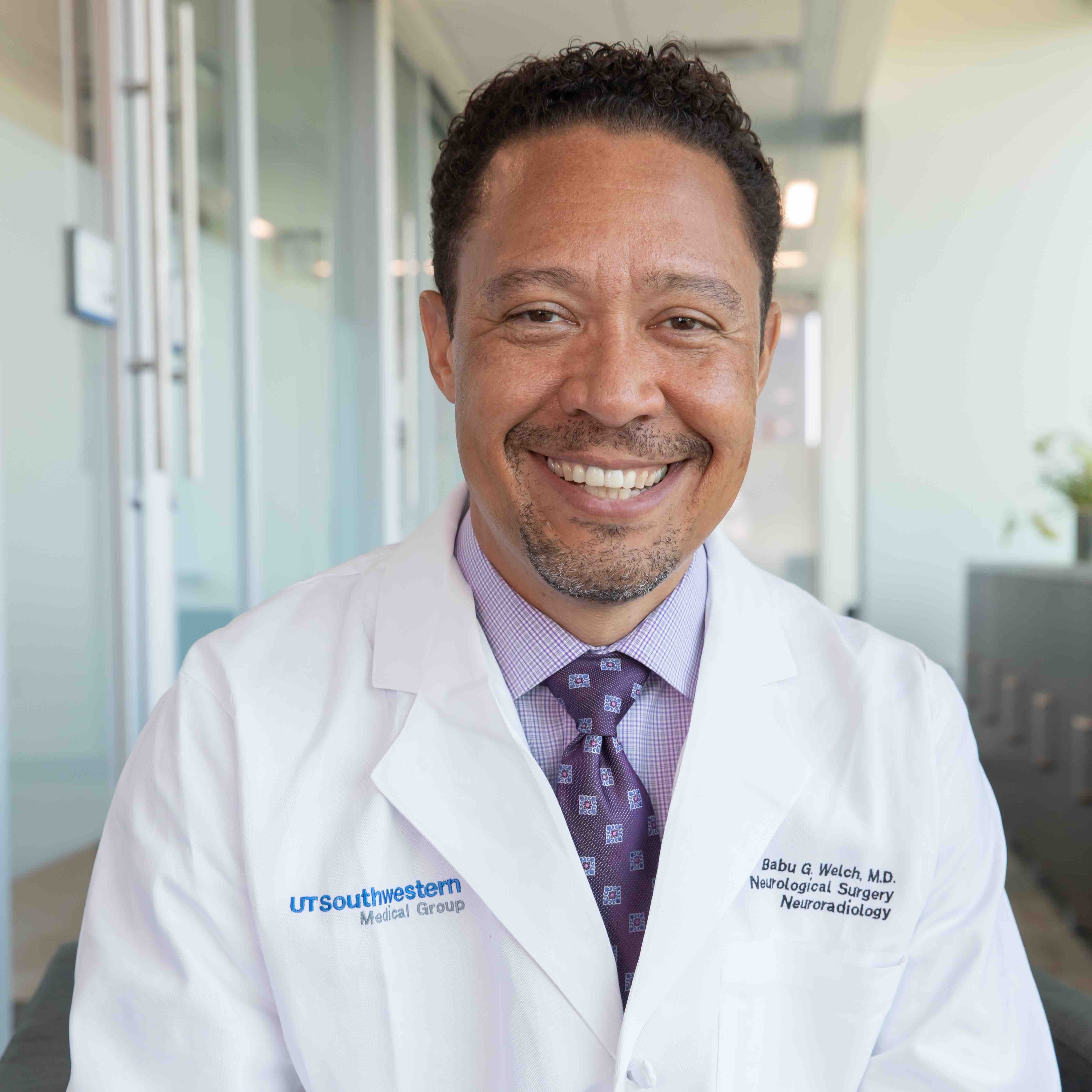
Babu Welch, M.D.
Then, during a second, eight-day hospital stay, doctors handed her a list of frightening possibilities, including multiple sclerosis, altered mental status, and cancer. That’s when Fowler insisted on being transferred more than 100 miles away to UT Southwestern’s Zale Lipshy Pavilion – William P. Clements Jr. University Hospital, following a friend’s recommendation.
Upon Fowler’s arrival at UT Southwestern’s Robert D. Rogers Advanced Comprehensive Stroke Center, her records went straight to Babu G. Welch, M.D., Professor of Neurological Surgery and Radiology, for review. Following additional testing, Dr. Welch confirmed a diagnosis: a brain arteriovenous malformation (AVM). Together, they mapped out a plan for surgery and rehabilitation.
The AVM Patient Profile
Fowler’s story reflects the confusion that often surrounds AVMs. Affecting just 1 in 15,000 people, these rare and unpredictable conditions frequently fly under the medical radar. While AVMs typically appear in young adults, they can also affect children – a population now served by UT Southwestern’s dedicated pediatric expertise. Compounding the challenge, many patients live for years without symptoms, completely unaware that they harbor a potentially dangerous lesion.
AVMs can present with seizures, hemorrhages, or persistent headaches, though some are discovered incidentally during brain scans for unrelated conditions such as chronic migraines. They can also appear as part of a larger syndrome, such as hereditary hemorrhagic telangiectasia (HHT). Because of its longstanding reputation for managing these complex malformations, UT Southwestern serves a primary referral center, drawing cases from across Texas and surrounding states to its multidisciplinary team of diagnostic and treatment specialists.
“We treat a large number – 40 to 60 – of AVMs every year and observe many more. Every case requires a multidisciplinary approach,” Dr. Welch said. “What really sets us apart is that we don’t begin treatment unless we have a clear, coordinated plan to see it all the way through.”
Evolution in Care
“In the 1990s and 2000s, endovascular therapy revolutionized how we approach AVMs. It allowed us to navigate through blood vessels and shut down abnormal connections from the inside,” Dr. Welch said. “It felt like a breakthrough, and many thought this was the cure for all AVMs, but the 2007 ARUBA trial forced us to recalibrate and redefine our approach when the results of an endovascular-heavy approach resulted in more patient harm than help.”
The meticulous yet controversial ARUBA study sparked great debate. Could treating AVMs with endovascular therapy be riskier than simple observation? Many specialists remain concerned that the aggressive, uncoordinated approach and results published in the ARUBA trial continue to discourage referrals of patients with AVMs to teams capable of treating the condition in a safer fashion. In a 2008 article responding to the findings, Dr. Welch advanced a broader understanding of AVMs as a complex disease that demands a multidisciplinary approach to determine the best plan for each patient, including endovascular, surgical, and radiosurgical options. The ARUBA study raised important questions, but it did not close the case on intervention.
At UT Southwestern, treating AVMs is a highly collaborative effort, uniting multiple specialties in comprehensive support for each patient.
Fowler experienced this collaboration firsthand. “Dr. Welch introduced the team, and my room was filled with white coats around my bed,” she said. “I remember saying, ‘There’s no way you’ll all be back tomorrow,’ but he smiled and said, ‘We’ll be here, won’t we?’ and in unison, they answered, ‘Yes, we’ll be here.’”
For unruptured AVMs, the team works together to determine whether observation, embolization, surgery, radiation, or a combination of therapies offers the safest and most effective path forward. For smaller AVMs, radiation oncologists can work with neurosurgeons to deliver highly targeted therapy with physicists, ensuring precise dosing and execution.
If an AVM ruptures, the team expands. Neurocritical care physicians stabilize patients in the ICU. Neurosurgeons and neurointerventional specialists address the source of bleeding, and neurology teams help with seizure management if necessary. Ultimately, rehabilitation specialists help patients recover function after stroke. From acute intervention to long-term recovery, Fowler’s journey illustrates the power of a medical community united to save lives and restore hope.
Continued Improvements
The most impressive improvements in AVM care are related to radiological imaging and endovascular techniques. Radiology is no longer just diagnostic: It is central to both risk assessment and treatment.
Functional imaging allows teams to determine how close an AVM is to critical brain centers, such as speech or movement, and to track whether those functions have shifted to other regions. Diffusion tract imaging maps motor pathways, sensory tracts, and visual fibers. Combined with advanced angiographic imaging, these tools help evaluate risk more precisely than ever before. Today, UT Southwestern has radiologists and neurologists who not only interpret detailed scans but also work with the neurosurgical team to perform endovascular procedures that directly treat AVMs.
If surgery is required, operating theaters called “hybrid suites” put everything the surgeon needs – surgical and radiology equipment – in one spot to decrease patient movement. UT Southwestern has used hybrid operating suites for surgical treatment of AVMs since 2007. Many major centers around the country have relied on this expertise to build their own facilities.
Together, these advancements influence decision-making and the refinement of best practices, which are shared at a longstanding multidisciplinary vascular conference. Keenly aware of how much the field has evolved since her 2012 diagnosis, Fowler recently shared her story at a regional neuroscience conference. There, she experienced a profound, full-circle, and emotional moment when she reunited with Dr. Welch, who saved her life more than a decade earlier.
“I follow the advances in technology and promote the earlier diagnosing of AVMs to get treatment plans shored up and implemented because, as they always say with stroke, time is brain, and with any type of neurological dysfunction you have to get on it,” Fowler said.
Living with AVMs
Fortunately, many patients with AVMs go on to live normal, active lives. Managing the condition typically relies on universal wellness strategies: maintaining blood pressure control, eating a healthy diet, and observing standard precautions if seizures are a factor. At UT Southwestern, the cerebrovascular team goes a step further, deliberately addressing both the biology of the lesion and the psychological mindset of the person living with it.
“The greater challenge for many patients is the ‘what if.’ Living with an untreated AVM can create anxiety about bleeding risk, even when that risk is low,” said Dr. Welch. “Some patients are comfortable with monitoring. Others find the uncertainty overwhelming, and that psychological burden can factor into treatment decisions.”
For patients undergoing observation, imaging scans are spaced out over years if the AVM remains stable. Patients are educated about symptoms that should prompt urgent evaluation, such as sudden severe headache, new neurological symptoms, or changes in seizure patterns. Over time, some patients become comfortable and simply check in periodically.
For those who undergo treatment, follow-up is structured. After radiosurgery, the effect can take up to five years as the vessels gradually thicken and close off. Imaging tracks that evolution to determine whether the AVM has been destroyed or if further treatment is needed. As in Fowler’s case, surgical treatment may be the best choice, and recovery following surgery might involve neurocritical care, rehabilitation, and sometimes the support of a neuropsychological team.
“There is no single pathway,” Dr Welch explained. “Treatment decisions depend on the AVM’s size and location, its relationship to critical brain structures, how the patient presented, and how they are tolerating the diagnosis emotionally. Care is individualized, and long-term personal partnership with an experienced team is essential.”
With the right care and continued understanding of AVMs, most patients can expect to live normal, productive lives.
Long View, Lasting Results
“We take pride in being a longstanding referral center for AVMs. There’s a deep culture here – built over decades – around caring for patients with these complex lesions,” Dr. Welch said. “In every specialty involved, there are senior physicians who have been here for more than 20 years.”
Neurosurgery, interventional radiology, radiation oncology, neurology, neurocritical care, and neurorehabilitation – each area has experienced anchors who have seen the field evolve and have cared for these patients throughout their journey, not just a moment in time. That continuity matters.
“We’re following patients for years, sometimes decades,” Dr. Welch noted. “There’s institutional memory here – and a shared commitment to seeing patients through treatment, recovery, and life beyond their AVM.”
This long view strengthens care so that every case adds to what teams know, every follow-up deepens understanding, and every recovery helps shape how the next patient is treated.